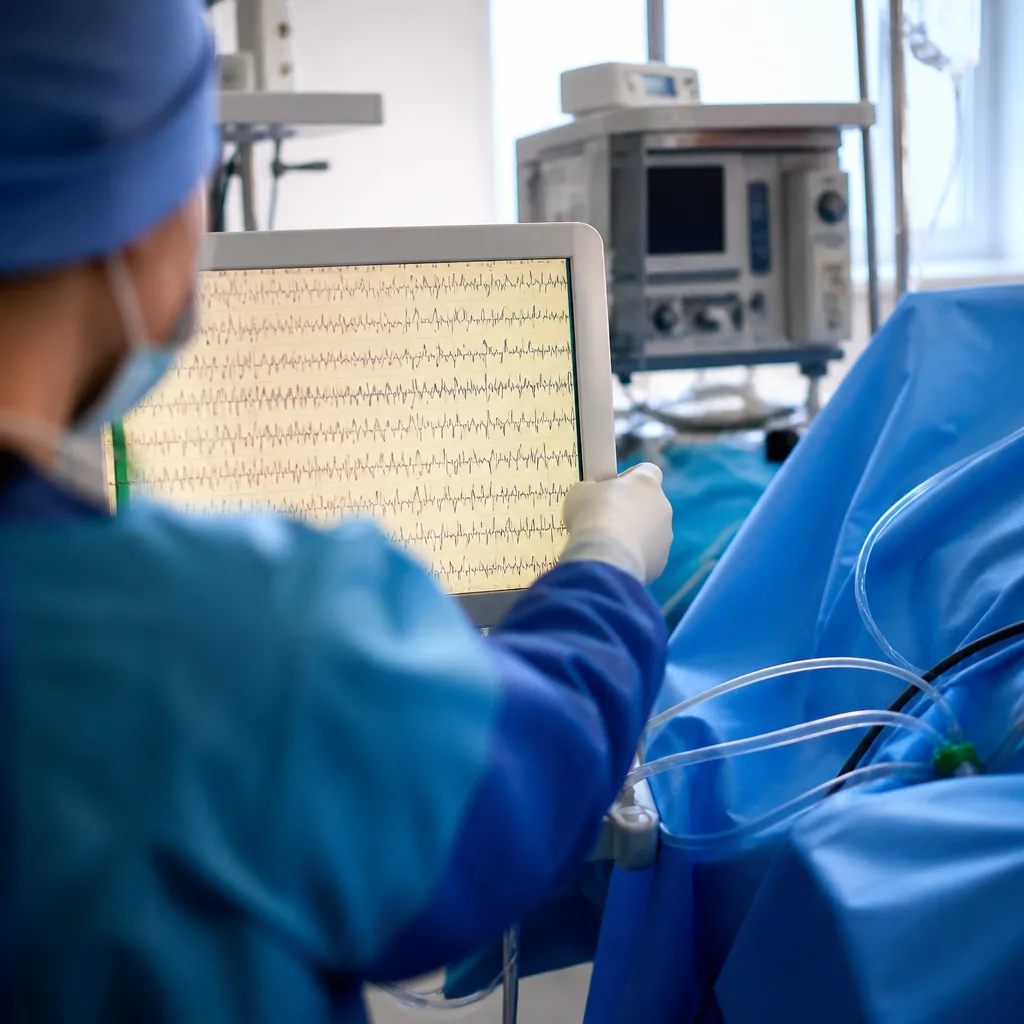
Anaesthetic drugs render people unconscious by changing how brain regions communicate, and electroencephalography (EEG) reveals predictable shifts in electrical activity as patients lapse into anaesthesia-induced unconsciousness, according to recent reporting of neuroscience and clinical work.
Anaesthesia as a research tool
Although anaesthesia is administered every day in operating theatres to allow surgery and manage pain, clinicians have not routinely monitored the brain itself during induction. In the past decade, neuroscientists interested in altered states of consciousness have increasingly used anaesthesia as an experimental tool to study how consciousness arises and fades. That shift has brought clinical and research communities closer, with potential benefits for perioperative brain health, particularly in older patients who are at greater risk of post‑operative cognitive problems.
George Mashour, a neuroanaesthesiologist at the University of Michigan whose work combines keeping patients unconscious during neurosurgery with studying brain activity under drugs, described the gap between practice and knowledge as “the central irony” of anaesthesiology. Mashour and colleagues track patients’ brain activity under anaesthesia with the objective of reducing adverse cognitive outcomes, an aim reflected in the 2018 American Society of Anaesthesiologists Brain Health Initiative report on perioperative brain health.
What EEG shows
EEG records the electrochemical interactions between communicating neurons through electrodes placed on the scalp and displays them as waveforms. As anaesthesia takes hold, the EEG signal typically changes in a characteristic way: waves tend to slow down and increase in amplitude. These alterations reflect disruption of the networks that sustain wakefulness and awareness.
| Brain region | Observed effect under anaesthesia |
|---|---|
| Cortex | Disrupted communication across cortical networks; cortical neuron inhibition |
| Thalamus and brainstem | Dampened connectivity between cortex, thalamus and brainstem |
One commonly used intravenous agent, propofol, is reported to enhance inhibition of cortical neurons and to interfere with cortical communication. The combined effect is a breakdown in the coordinated signalling that supports conscious experience.
Clinical implications and remaining questions
Using EEG to follow these sequence changes gives clinicians and researchers a window into the brain’s transition into and out of anaesthesia. Yet routine brain monitoring during anaesthesia is not universal. Advocates argue that better intraoperative brain monitoring could help tailor anaesthetic dosing and potentially reduce the incidence of post‑operative delirium and longer‑term cognitive decline in vulnerable populations.
At the same time, scientists caution that while the broad patterns are reproducible, the precise mechanisms that produce unconsciousness under different anaesthetics are not fully understood. Anaesthesia affects multiple molecular targets and network interactions, so findings from EEG and other measures must be interpreted carefully.
- EEG provides a reproducible signature of the brain’s shift into anaesthesia — slower, higher‑amplitude waves.
- Propofol acts principally by increasing cortical inhibition and disrupting cortical and thalamocortical communication.
- Clinical monitoring lag: despite advances, brain monitoring is not yet routine during all anaesthetic care.
“It’s the central irony,” said George Mashour, describing how anaesthesiology relies almost entirely on brain‑acting drugs without routinely measuring the brain during care.
Research that treats anaesthesia as a controlled perturbation of brain function is likely to continue informing both basic science about consciousness and practical approaches to perioperative brain health. For clinicians, the challenge is translating group‑level EEG patterns into personalised care that reduces harm while preserving the indispensable role of anaesthesia in modern surgery.