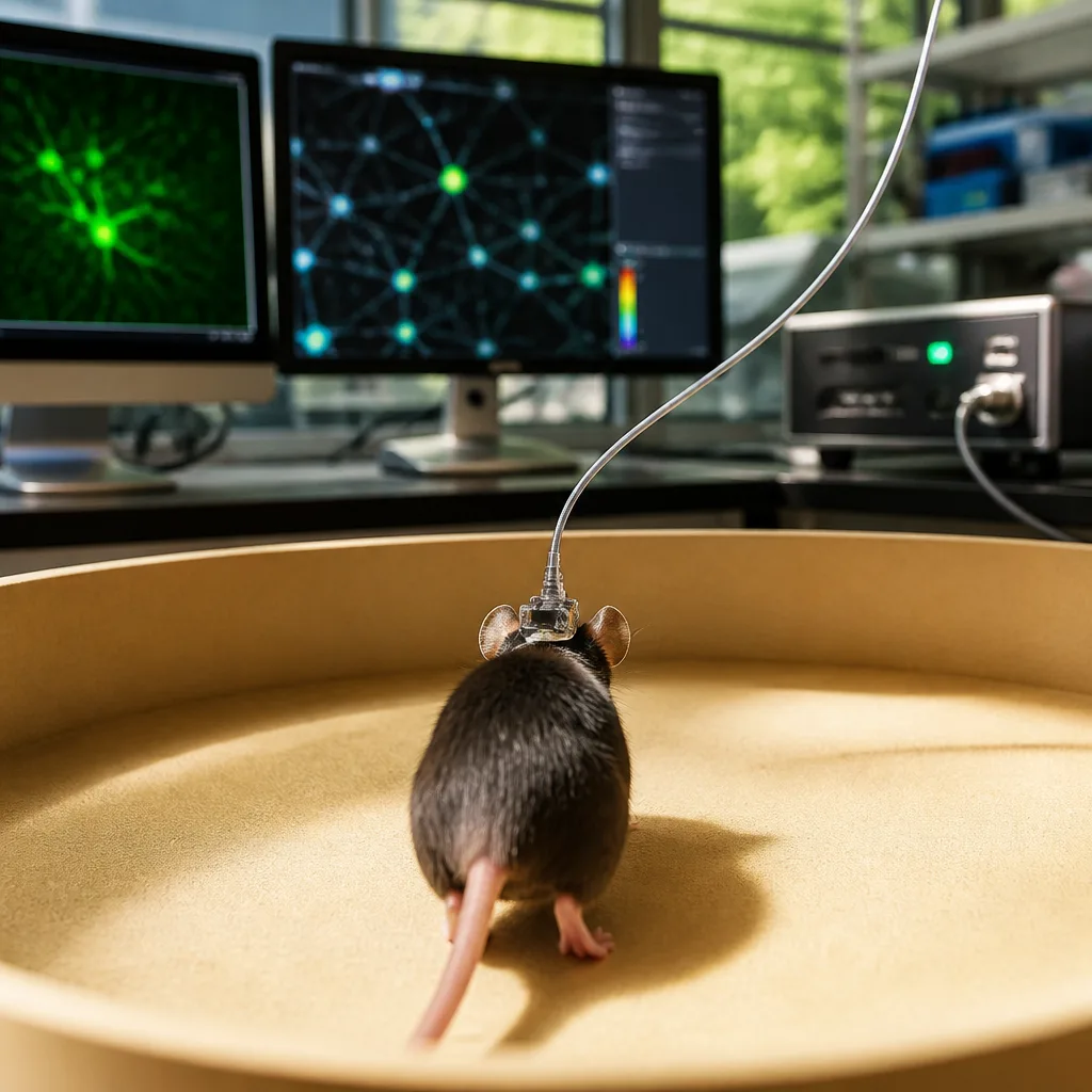
Researchers at the University of Colorado Anschutz and the University of Colorado Boulder have unveiled a lightweight, head-mounted microscope that can both record high-resolution activity from individual brain cells and trigger those same cells with light while animals move freely.
Dual-readout and control in a single device
The device, named Opto2P-FCM, combines two-photon imaging with patterned photostimulation in a compact, fibre-coupled package. Two-photon techniques allow for deeper penetration into brain tissue and finer resolution than many conventional miniature microscopes; pairing that capability with targeted optogenetic stimulation gives researchers simultaneous readout and control of specific neurons during natural behaviour.
Developers say the instrument is unusually light: the team describes engineering the system at roughly five grams, allowing it to be mounted on an animal's head without substantially impeding movement. The work appears in the journal Optica.
“These miniature microscopes have been increasingly used to study the neural basis of behavior in freely moving animals,” said Emily Gibson, Ph.D., associate professor of biomedical engineering at CU Anschutz and the study's senior author. “This new microscope allows deeper penetration and higher-resolution images of the brain. Importantly, it can also excite select neurons using optogenetics.”
Why the combination matters
Until now, many head-mounted microscopes could either image neural activity or, separately, deliver light for optogenetic manipulation. Opto2P-FCM integrates both functions, enabling experiments that directly link the activity of identified neurons to ongoing behaviour by turning those neurons on or off while simultaneously tracking network responses.
Juliet Gopinath, Ph.D., a co-author and professor at CU Boulder, called the instrument a potential catalyst for future discoveries, noting the significance of having “state-of-the-art” readout and photostimulation in a single platform. Co-first author Mo Zohrabi, Ph.D., described the development as overcoming an engineering challenge the team once thought improbable.
Potential research impacts
By permitting targeted manipulation and observation of individual cells in untethered animals, the technology opens avenues for experiments that probe how particular neurons contribute to decision-making, movement, learning and other behaviours in environments that better mirror natural conditions. The authors suggest the instrument could help deepen mechanistic understanding of disorders that arise from circuit dysfunction.
- Imaging mode: two-photon, fibre-coupled for deeper tissue access and higher spatial resolution.
- Stimulation: patterned photostimulation allowing selective excitation of chosen neurons via optogenetics.
- Form factor: head-mounted and lightweight (about five grams) to permit freely moving animal experiments.
| Feature | Capability |
|---|---|
| Imaging modality | Two-photon fibre-coupled imaging |
| Stimulation | Patterned optogenetic photostimulation |
| Weight | ~5 grams (head-mounted) |
Limits and next steps
The report focuses on the design and optical performance of the Opto2P-FCM, demonstrating that simultaneous imaging and stimulation are technically achievable in a miniaturised format. Details on long-term deployment in varied behavioural paradigms, the breadth of brain regions accessible, and adaptation to other species or experimental preparations will emerge as other labs test and adopt the system.
For the neuroscience community, tools that merge observation and manipulation are increasingly valued because they enable causal tests of neural circuit function. The Opto2P-FCM adds to a growing toolbox aimed at bridging cellular-scale measurements with natural behaviour — an essential step toward translating basic circuit insights into understanding and, ultimately, treatments for neurological conditions.
Authors on the paper highlight the technical achievement and the scientific opportunities it creates, while acknowledging that broader uptake will depend on reproducibility, ease of use and integration with genetic and behavioural methods already used in laboratories worldwide.
The research was published in Optica, and the team emphasises the instrument’s potential to accelerate studies into how the brain controls behaviour and how disorders of neural circuits arise.