
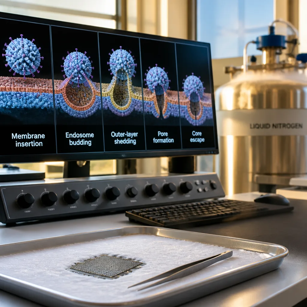
Researchers have used cryo‑electron tomography to record, at near‑molecular resolution, how a non‑enveloped rotavirus penetrates a host cell, tracing a sequence of structural events that end with the viral core entering the cell cytoplasm and beginning replication. The work, led by postdoctoral fellow Marilina de Soto at Harvard Medical School, was reported in the journal Science on 10 September.
What the images show
Cryo‑electron tomography is a three‑dimensional imaging method in which biological material is rapidly frozen at cryogenic temperatures and imaged from multiple angles. The technique enabled the team to visualise the interaction between a rhesus rotavirus and the cell membrane in situ, at resolutions below 1 nanometre.
The dataset reveals a clear, repeated sequence of events during viral entry:
- Attachment and membrane insertion: virus surface spikes insert into the host membrane;
- Membrane wrapping and endocytosis: those spikes induce the membrane to curve and envelop the particle, forming an endosomal compartment;
- Outer layer shedding: after internalisation, the virus loses its outer protein layer;
- Pore formation: the same surface proteins then perforate the endosomal membrane;
- Core escape and initiation of replication: the infectious core translocates through these pores into the cytoplasm, where it uses internal enzymes to make messenger RNA and begin replication.
"Cryo‑electron tomography allowed us to visualize the entire process by"
The quoted fragment is from the authors’ report in Science, emphasising that the imaging method captured the complete entry sequence for a non‑enveloped virus — a class of viruses that lack a surrounding lipid membrane and therefore cannot fuse with cell membranes the way enveloped viruses do.
Why this matters
Viruses fall into two broad structural categories: those with a lipid envelope and those without. Enveloped viruses typically fuse that envelope with the host membrane to enter a cell. Non‑enveloped viruses, such as rotavirus, must rely on alternate mechanical and protein‑mediated strategies to cross the membrane barrier. Direct visualisation of these steps for rotavirus clarifies how surface proteins perform multiple, distinct mechanical roles during entry.
The study provides a structural framework for understanding how the infectious core of rotavirus — the transcriptionally active particle that produces viral mRNA — reaches the cytoplasm intact. This is a critical stage in infection, because the core carries enzymes that immediately begin viral gene expression once released from the endosome.
Implications and caveats
The findings refine fundamental knowledge of viral entry and may guide future laboratory work aimed at disrupting specific stages of the process. For example, blocking the membrane insertion or pore‑forming actions of surface proteins could, in principle, prevent the core’s release into the cytoplasm.
However, the report presents observational structural data rather than therapeutic trials. The images document what happens at high resolution; they do not by themselves demonstrate how to interrupt the process in a clinical setting. Further biochemical and functional studies are needed to test interventions that target the newly described steps.
Rotavirus remains a significant cause of severe gastroenteritis in infants and young children worldwide. Understanding its entry strategy does not immediately change clinical practice, but it strengthens the scientific basis for antiviral and vaccine research by identifying precise mechanical actions that might be susceptible to inhibition.
| Stage | Description |
|---|---|
| Attachment | Spike proteins contact and insert into the plasma membrane |
| Endocytosis | Membrane invaginates to form an endosome around the virion |
| Shedding | Outer protein layer is removed inside the endosome |
| Pore formation | Surface proteins create pores in the endosomal membrane |
| Core escape | Infectious core passes into cytoplasm and begins mRNA synthesis |
The study exemplifies how advanced imaging techniques can capture dynamic viral processes in near‑native conditions. As cryo‑electron tomography and related methods become more widely used, comparable mechanistic pictures for other non‑enveloped viruses may emerge, helping to build a broader, structural understanding of viral entry strategies across pathogen families.


