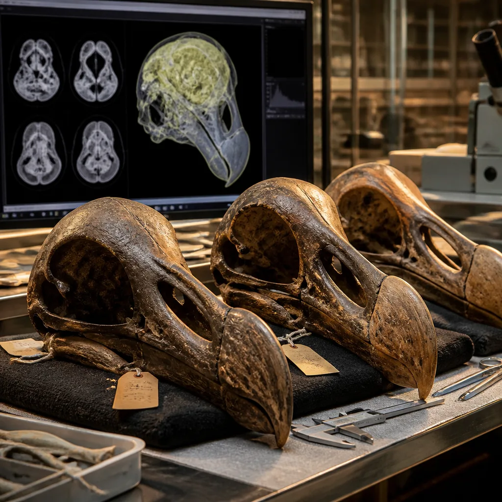
The long-standing image of the dodo as a comical, foolish bird has been challenged by a new comparative study that used high-resolution CT imaging to reconstruct the internal shape of the extinct species’ braincase. Working with the only three largely complete dodo skulls known to science, researchers found features consistent with a well-developed sense of smell and forward-oriented, owl-like eyes, rather than a disproportionately small brain.
How the reconstruction was done
An international team led from the University of Lethbridge in Canada obtained access to three rare skulls: two nearly complete specimens and a third damaged but amenable to digital reconstruction. The specimens are held in major museum collections in Europe and the United States. High-resolution CT scans were used to image the internal cavities of the skulls — the braincase that once housed the brain — enabling virtual reconstructions of the relative sizes of distinct neural regions.
The researchers did not examine the soft tissue of a living brain; instead they inferred the proportions of sensory and cognitive centres by measuring the spaces within the skull where those brain regions would have sat. To place the dodo’s endocranial anatomy in context, the team compared the results with scans of the Rodrigues Solitaire, the dodo’s nearest extinct relative, and with skulls from eight living species of pigeons and doves.
Findings and interpretation
The study reports two notable departures from the popular picture of the dodo. First, the volume of the olfactory region — the part of the brain associated with smell — was larger than might be expected for a bird of its size. Second, the orientation and inferred sighting geometry are consistent with forward-facing, “owl-like” eyes, implying adaptations for binocular vision and possibly for seeing in low-light conditions.
Importantly, the dodo’s overall brain size was not found to be abnormally small for a bird of its body dimensions. In other words, the species’ reputation for stupidity is not supported by the endocranial evidence now available. Instead, the reconstructed anatomy suggests a bird adapted to detect odours and to operate in dimly lit environments on Mauritius.
- Specimens studied: three largely complete dodo skulls from museum collections.
- Comparisons: Rodrigues Solitaire and eight living pigeons and doves.
- Key anatomical signals: enlarged olfactory centre; forward-facing ocular orientation; brain size within expected range.
| Specimen | Repository |
|---|---|
| Dodo skull (nearly complete) | Natural History Museum of Denmark |
| Dodo skull (nearly complete) | Oxford University Museum of Natural History |
| Dodo skull (damaged, reconstructable) | Smithsonian Institution |
Context and consequences
The dodo (Raphus cucullatus) has been extinct since the late 17th century and much of what the public knows about it derives from sailors’ accounts and artistic depictions rather than direct anatomical study. Museum collections therefore provide the only route to test long-standing assumptions. By applying modern imaging to these rare remains, the authors provide a firmer anatomical basis for interpreting how the bird sensed and interacted with its environment.
That reinterpretation carries cultural weight. The dodo’s name has become shorthand for ineptitude in many languages and popular culture. Demonstrating that the bird was not neurologically diminutive but instead equipped with sensory specialisations invites a reassessment of that caricature. From a scientific standpoint, the study underlines how extinction narratives based on limited historical observation can be revised when museum specimens are re-examined with modern techniques.
Methodologically, the work also highlights the value of comparative endocranial studies: by measuring relative brain-region sizes across related taxa, researchers can infer sensory priorities and ecological niches even in species known only from bones. The approach is particularly powerful for island endemics such as the dodo, whose ecology was shaped by isolated conditions and unique selective pressures.
Further research could expand the taxonomic sample of extant comparators or explore how the dodo’s cranial anatomy relates to ecological data from subfossil deposits on Mauritius. For now, the principal finding is clear: available evidence points to a bird adapted to detect smell and to operate with forward-facing vision, not to the hapless creature of popular imagination.