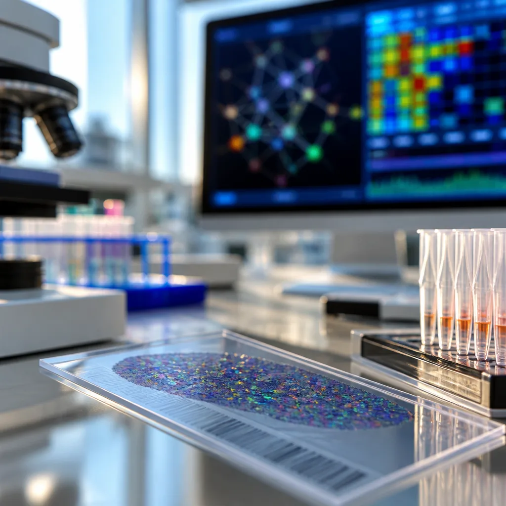
Spatial multiomics techniques are now enabling scientists to measure the transcriptome, proteome and epigenome from the same tissue section, allowing direct comparison of RNA levels, protein abundance and chromatin state in their native cellular neighbourhoods. The shift from single‑layer mapping to joint profiling marks a major advance for spatial biology and is already reshaping both laboratory practice and computational tool development.
What spatial multiomics delivers
Historically, different molecular layers had to be assayed separately or on serial sections, forcing researchers to stitch datasets together and infer relationships across experiments. Spatial multiomics removes that dislocation by co‑profiling multiple modalities from the same physical sample. That means a researcher can now investigate how chromatin accessibility corresponds to gene expression and how those transcriptional patterns translate into protein distribution within the precise architecture of tissue.
Sequencing‑based co‑detection approaches are pushing resolution and throughput. One method highlighted by recent reviews can map the whole transcriptome while simultaneously detecting surface proteins, reporting capability for up to 273 proteins alongside RNA at near‑single‑cell spatial resolution. Beyond two‑dimensional sections, platforms are beginning to extend co‑profiling into three dimensions and volumetric specimens, and live imaging efforts aim to add a temporal axis.
Computational headaches and emerging solutions
Combining different molecular layers in space introduces complex integration problems. Modalities vary in spatial resolution, sparsity, dynamic range and data type. Aligning such heterogeneous measurements and extracting coherent biological signals is far from trivial. As the field scales, developers are turning to advanced machine learning frameworks; graph neural networks are among the approaches being used to represent tissue architecture and harmonise signals across layers.
- Diverse data properties: gene expression, protein counts and chromatin marks behave differently and require tailored preprocessing.
- Spatial alignment: measurements must be co‑localised precisely to preserve tissue context.
- Scale: 3D and temporal initiatives increase data size and complexity.
These challenges are driving a new generation of computational tools that do more than visualise layers; they integrate regulatory relationships and provide testable hypotheses about molecular mechanisms in situ.
Early impact and directions
Adopters of spatial multiomics are already reporting insights across several fields. Tumour biology benefits from simultaneous maps of gene regulation and protein distribution, revealing the microenvironmental interactions that shape therapeutic response. Neuroscience gains a way to place transcriptional programmes and epigenetic states within defined circuits. Developmental biology can trace how regulatory layers coordinate during morphogenesis when read directly in tissue.
As methods mature, expectations are high but realistic: spatial multiomics does not replace single‑modality experiments but complements them, offering a richer, physically grounded picture of cellular state. Laboratories will need both wet‑lab protocols capable of preserving multiple molecular targets and computational pipelines that reconcile their different characteristics.
For industry and funding bodies, the message is clear — investment is required not only in instruments and reagents but also in scalable software, data standards and training. Without harmonised approaches, the promise of multi‑layer spatial maps could be blunted by incompatible datasets and reproducibility gaps.
In practical terms, researchers aiming to adopt these techniques should plan for cross‑disciplinary teams: biologists, technologists and computational scientists working together to validate measurements and interpret integrated maps. The coming years will show whether spatial multiomics becomes routine in research labs and clinical translation, but the technology has already altered how scientists conceptualise molecular biology in context.
| Feature | Current capability |
|---|---|
| Proteins detected alongside transcriptome | Up to 273 |
| Spatial dimension | 2D sections; emerging 3D/volumetric and live imaging |
| Computational focus | Alignment across resolutions, sparsity handling, graph neural networks |