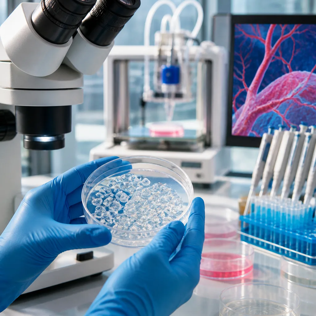
Researchers at Penn State have demonstrated a method to 3D print tiny clusters of living cells — known as spheroids — that are engineered to support the regeneration of bone tissue and the formation of new blood vessels. The work, reported in the Chemical Engineering Journal and validated in lab experiments and mouse models, outlines an approach to programme undifferentiated stem cells so they mature into multicellular structures useful for repairing damaged bone.
Programme cell behaviour rather than rely on scaffolds
Bioprinting commonly places cells inside a hydrogel scaffold that provides a three-dimensional matrix for cells to grow. The Penn State team took a different tack: by introducing distinct strands of genetic information into commercially sourced stem cells prior to printing, they created spheroids whose internal composition encourages both bone formation and vascularisation as the cluster develops. That combination is crucial because new blood vessels are needed to sustain regenerated tissue.
"Everything in your body is made up of a combination of different cell types, meaning we have to find a way of differentiating cells from one another as they mature when forming these tissues," said Daniel Hayes, co-corresponding author and head of Penn State's Department of Biomedical Engineering.
The researchers say the method is intended to build spheroids that can be used to reconstruct complex cellular architectures. In laboratory tests and in mice, the engineered spheroids showed improved capacity to aid bone healing and supported the successful formation of vasculature within the generated tissue.
Potential uses and current limits
The team highlights two clear applications. First, such spheroids could become a component of regenerative medicine strategies for severe trauma or infections that damage bone. Second, they offer a platform for producing accurate biological models for experimental drug testing — a potentially valuable tool for evaluating candidate treatments in structures that better mimic real tissue.
But the authors caution that translating the technique into clinical therapies is not straightforward. The central challenge is replicating the diversity of cell types present in real organs and ensuring they differentiate appropriately as the structure matures. The research demonstrates a proof of concept: that supplying different genetic instructions to the starting stem cells can produce clusters predisposed to desired functions.
- What was achieved: Bioprinted spheroids engineered through genetic instructions that support bone regeneration and vascular growth.
- Evidence: Results verified in laboratory experiments and in mouse models; findings published in Chemical Engineering Journal.
- Applications: Regenerative medicine for bone injuries and improved in vitro models for drug testing.
| Aspect | Implication |
|---|---|
| Spheroid programming | Enables targeted differentiation and function without relying solely on scaffold properties |
| Vascularisation | Essential for survival and integration of regenerated tissue; demonstrated in mouse models |
| Translational challenge | Complexity of recreating multiple cell types and ensuring correct maturation |
The lead investigators include Daniel Hayes, who holds the Dorothy Foehr Huck and J. Lloyd Huck Chair in Nanotherapeutics and Regenerative Medicine and is head of Penn State's Department of Biomedical Engineering. The research team combined expertise in engineering and chemistry to develop and test the approach.
While the work stops short of clinical application, it advances a growing body of research that seeks to move beyond simple cell-laden hydrogels towards printed constructs whose cellular composition and behaviour are pre-programmed. If further studies confirm safety and efficacy, the method could become a building block for future therapies aimed at reconstructing damaged bone and other complex tissues.
For now, the contribution is an important technical step: demonstrating that bioprinting can produce spheroids optimised for both tissue formation and vascular support, rather than relying solely on the passive environment of a scaffold to drive cell differentiation and organisation.